summary: Researchers have discovered a unique feature of communication networks in the human brain: the transmission of information along multiple parallel pathways, a feature that has not been observed in macaques or mice.
This finding emerged from a study that used diffusion and fMRI data, as well as information and graph theory. The team mapped “brain movement” to compare signal transmission in the brains of different mammals.
Their research suggests that these parallel pathways in humans may contribute to our advanced cognitive abilities and could have implications for understanding brain evolution and potential medical applications.
Key facts:
- The EPFL study found that human brains uniquely transmit information through multiple parallel pathways, unlike macaques and mice.
- This discovery was made using a new combination of diffusion MRI, functional MRI, information theory, and graph theory.
- The research suggests that these parallel pathways could contribute to higher cognitive functions and offer new insights into brain plasticity and neurorehabilitation.
source: EPFL
In a study that compared communication networks in the human brain with those in macaques and mice, EPFL researchers found that only human brains transmit information along multiple parallel pathways, leading to new insights into mammalian evolution.
When describing brain communication networks, Alessandra Griffa, a postdoctoral researcher at EPFL, likes to use travel metaphors. Brain signals are sent from source to target, creating a multisynaptic pathway that intersects multiple brain regions “like a road with many stops along the way.”

She explains that structural communication pathways of the brain have already been observed based on networks (“routes”) of nerve fibers. But as a scientist at the Medical Image Processing Laboratory (MIP:Lab) at EPFL’s Faculty of Engineering, and a research coordinator at CHUV’s Leenaards Memory Centre, Griffa wanted to follow patterns of information transmission to learn how messages are sent and received. In a study recently published in nature communications, She worked with MIP:Lab head Dimitri van de Wiel and SNSF Ambizione Fellow Enrico Amico to create “brain movement maps” that could be compared between humans and other mammals.
To achieve this, the researchers used open-source diffusion (DWI) and functional magnetic resonance imaging (fMRI) data from humans, macaques and mice, which were collected while the subjects were awake and at rest.
DWI scans allowed scientists to reconstruct brain “road maps,” and fMRI scans allowed them to see different areas of the brain lighting up along each “road,” suggesting that these pathways were transmitting neural information.
They analyzed multimodal MRI data using information theory and graph theory, and Greva says it was this new combination of methods that led to new insights.
“What’s new in our study is the use of multimodal data in a single model that combines two branches of mathematics: graph theory, which describes multi-synaptic ‘roadmaps’; and information theory, which defines the transfer of information (or ‘traffic’) across roads.”
“The basic principle is that messages passed from source to target remain unchanged or deteriorate further at each stop along the way, like the game of telephone we played as children.”
The researchers’ approach revealed that in non-human brains, information is sent along a single “route”, whereas in humans, there were multiple parallel paths between the same source and target. Furthermore, these parallel tracks were as unique as fingerprints and could be used to identify individuals.
“Such parallel processing has been hypothesized in human brains, but has never before been observed at the whole-brain level,” Greva summarizes.
Possible insights into evolution and medicine
The beauty of the researchers’ model, Griffa says, lies in its simplicity, and that it inspires new perspectives and research avenues in evolution and computational neuroscience. For example, the findings could be linked to the expansion of human brain size over time, giving rise to more complex connectivity patterns.
“We can assume that these parallel information flows allow multiple representations of reality, and the ability to perform abstract functions specific to humans.”
She adds that although this hypothesis is just speculation, as in… Nature Communications The study did not include any testing of people’s arithmetic or cognitive ability, and these are questions that she would like to explore in the future.
“We looked at how information is transmitted, so an interesting next step will be to model more complex processes to study how information is combined and processed in the brain to create something new.”
As a memory and cognition researcher, she is particularly interested in using the model developed in the study to see whether parallel information transfer can confer plasticity to brain networks, perhaps playing a role in neurorehabilitation after brain injury, or in the prevention of cognitive decline. In diseases of aging.
“Some people age healthily, while others experience cognitive decline, so we would like to see whether there is a relationship between this difference and the presence of parallel information flows, and whether they can be trained to compensate for neurodegenerative processes.”
About this neuroscience research news
author: Celia Lauterbacher
source: EPFL
communication: Celia Lauterbacher – EPFL
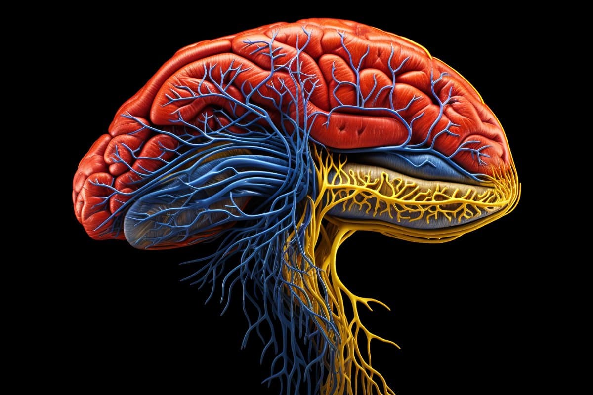
picture: Image credited to Neuroscience News
Original search: Open access.
“Evidence for increased parallel information transfer in human brain networks compared to male macaques and mice“By Dimitri van de Wiel et al. Nature Communications
a summary
Evidence for increased parallel information transfer in human brain networks compared to male macaques and mice
Brain communication, defined as the transmission of information via white matter connections, is the basis of the brain’s computational capabilities that encompass almost all aspects of behavior: from sensory perception common to mammalian species, to complex cognitive functions in humans.
How have communication strategies in large brain networks adapted over evolution to accomplish increasingly complex functions?
By applying a graph approach and information theory to evaluate information-related pathways in the brains of male mice, macaques, and humans, we demonstrate the brain connectivity gap between selective information transfer in non-human mammals, where brain regions share information through individual multi-synaptic pathways. Parallel information transfer in humans, where regions share information through multiple parallel paths. In humans, parallel transport serves as a major link between unilateral and transmodal systems.
The layout of information-related pathways is unique to individuals across different mammalian species, suggesting specificity of information routing structure at the individual level.
Our work provides evidence that different connectivity patterns are linked to the evolution of brain networks in mammals.

“Beer aficionado. Gamer. Alcohol fanatic. Evil food trailblazer. Avid bacon maven.”
